Showing posts with label Introduction to biochemistry laboratory. Show all posts

Saturday, January 5, 2013

What is Dry chemistry ?

Dry Chemistry:

The main principle of dry chemistry is based upon the reflectance spectrophotometry.

Reflectance spectrophotometry measures the reflectance of materials. Reflectance measurements are of great value in providing a reference standard for the comparison of the colour of different samples.
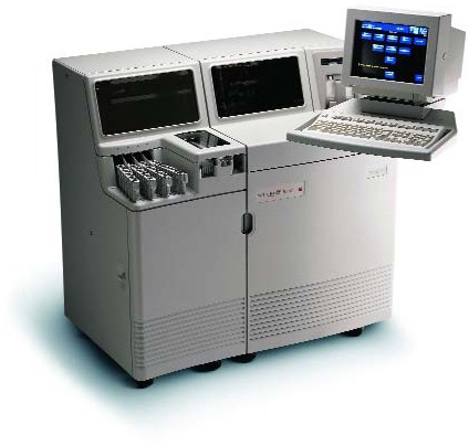
Fig. Vitros 250 Dry chemistry analyzer

Reflectance measurements are made using both diffuse and specularly reflected light.In diffuse reflectance, light is scattered in all directions from the sample.Provided that this scattered light can be collected onto an optical detector, the surface reflectance may be measured either at a given wavelength, or by performing a scan over a range of wavelengths. Such a wavelength scan can then be used to characterise colour.

A reflectance spectrophotometer is similar to a standard UV/Visible spectrophotometer.It should have a bandwidth narrow enough to provide well resolved visible spectra yet wide enough to provide a good energy level for diffuse reflectance measurements. The reflectance spectrophotometer must also have optics and electronics systems of high sensitivity, and should be able to physically accommodate reflectance and transmission accessories. The adapted spectrophotometer must be able to make measurements both at selected fixed wavelengths or perform scans over the complete wavelength range.

In dry chemistry, slides are dry, multilayered analytical elements coated on polyester supports. A small amount of patient sample is deposited onto the slide and evenly distributed to all of the layers. The spreading layer contains the appropriate substrate and other components needed for the reaction. The analyte in the sample catalyzes the reaction sequence to yield products which absorb light at wavelengths in various regions (340 – 680nm), diffuses into the underlying layer, and is monitored by reflectance spectrophotometry. The test types are colorimetric, enzymatic end point, two-point or multi-point rate, or potentiometric. The rate of change in reflection density in converted to enzymatic activity or the amount of colored complex formed is proportional to the analyte concentration in the sample.


Specimen requirement:

2 ml of serum collected in a red top tube with a serum separator (gel barrier). Centrifuge the specimen after it has clotted to prevent hemolysis. Send to the lab at room temperature. If the blood is not sent to lab the same day it is drawn, centrifuge the specimen and refrigerate. Serum that is hemolyzed and/or lipemic may interfere with some chemistries and may be rejected.

Sunday, November 11, 2012

USE OF RADIACTIVE SUBSTANCE


Iodine-131 and phosphorus-32 are also used for therapy. Iodine-131 is used to treat the thyroid for cancers and other abnormal conditions such as hyperthyroidism (over-active thyroid). In a disease called Polycythemia Vera, an excess of red blood cells is produced in the bone marrow. Phosphorus-32 is used to control this excess. 

Radiolabelled Cr is used to assess GFR. Many Radioimmunoassays of hormones are used for diagnosis of endocrine disorders. Fluoro-deoxy glucose (FDG) incorporating F-18 with a half-life of just less than two hours is readily incorporated into the cell without being broken down, and is a good indicator of cell metabolism. Lead-212, with half-life of 10.6 hours, can be attached to monoclonal antibodies for cancer treatment.

A scintillator is a material, which exhibits scintillation—the property of luminescence when excited by ionizing radiation. Luminescent materials, when struck by an incoming particle, absorb its energy and scintillate, i.e., reemit the absorbed energy in the form of light. Sometimes, the excited state is metastable, so the relaxation back out of the excited state is delayed and can lead to delayed fluorescence or phosphorescence, also called after-glow.

A scintillation detector or scintillation counter is obtained when a scintillator is coupled to an electronic light sensor such as a photomultiplier tube (PMT) or a photodiode. PMTs absorb the light emitted by the scintillator and reemit it in the form of electrons via the photoelectric effect.
Scintillation counter measures the radioactivity. The principle types of scintillation detectors found in clinical chemistry laboratory are crystal scintillation detector and the organic liquid scintillation detector. In this process the absorbed energy produces a flash of light, rather than a pulse of current. Crystal scintillation detects gamma emitters like 15Cr, 131I, etc. Liquid scintillation detects beta emitters like tritium, 32P, 14C.

The sensor called scintillator consists of transparent crystal (made up of Na iodide that can absorb gamma ray) or organic liquid (liquid scintillation counting, contains aromatic solvents and fluor a scintillator) that fluoresces when struck by ionizing radiation. A sensitive photomultiplier tube measures the light from crystal. The PMT is attached to amplifier and other electronic equipment to count and quantify the amplitude of the signal.

In liquid scintillation counter beta particles emitted from the sample transfer energy to the solvent molecules, which in turn transfer their energy to the fluors; the excited fluor molecules dissipate the energy by emitting light. In this way, each beta emission (ideally) results in a pulse of light. Scintillation cocktails often contain additives that shift the wavelength of the emitted light to make it more easily detected. These generated light pulses, reach both photomultiplier tube which, are counted.

(Source: Wikipedia) 

ANTICOAGULANTS AND PRESERVATIVES FOR BLOOD


Chemical agents that prevent coagulation are routinely used when whole blood or plasma is required. Some of the commonly used anticoagulants are:

(1) Heparin         (2) Salts of Ethylene diamine tetra acetic acid (EDTA)
(3) Oxalates        (4) Sodium Fluoride 

Heparin

It is the most widely used anticoagulant and causes least interference with tests. It is available as sodium, potassium, lithium and ammonium salts. This anticoagulant accelerates the action antithrombin III which neutralizes thrombin and prevents conversion of fibrin form fibrinogen. 

It is used in 0.2mg/ml blood. Its disadvantage is high cost and short action, and produces blue background in blood smears. It is believed to inhibit the activity of acid phosphatase and affect binding of T3 and T4 to carrier proteins producing high free concentrations of these hormones.

EDTA

It is a chelating agent useful in hematology as it preserves the cellular component of blood. It is used as disodium, dipotassium or tripotassium salt. It is used as 1-2mg/ml blood. EDTA prevents coagulation by chelating calcium. 

Dry EDTA vials are used. It by chelating metallic cofactors, inhibits alkaline phosphatase, creatine kinase and other enzymes. It is not suitable to use in specimen for calcium and iron analysis.

Sodium fluoride

It is used when blood is collected for glucose estimations. It is a weak anticoagulant but is often added as preservative for blood glucose together with potassium oxalate as anticoagulant. It is effective at a concentration of 2mg/ml blood. It inhibits the enzyme enolase (sodium fluoride) thus inhibiting glycolysis. Na fluoride/K oxalates are mixed in the ratio 1:3.

Citrate

Sodium citrate solution at concentration 3.8g/dl in ratio or 1 part to 9 part of blood is widely used for coagulation studies because its effect is reversible by addition of calcium.

Oxalates

Sodium, potassium, ammonium, and lithium oxalates inhibit blood coagulation by forming insoluble complex with calcium. Potassium oxalate at concentration of 1-2 mg/ml of blood is widely used. Combined ammonium and/or potassium oxalate does not cause shrinkage of erythrocytes. It consists of three parts by weight of ammonium oxalate, which causes swelling of the erythrocytes, balanced by two parts of potassium oxalate which causes shrinkage. NH4+ & K+ oxalate mixture in the ratio of 3:2, and 2 mg / ml of blood is the required amount.


The disadvantage of the use of oxalate is the alteration of concentrations of plasma components.  Shrinkage of erythrocytes results from a water shift from the erythrocytes to plasma. This shift increases with increasing anticoagulant concentration, and if used in the same concentration on a weight basis, all anticoagulants will have this effect inversely proportional to their molecular weight.  Aside from the water shift there may be alteration of erythrocyte permeability, which may explain the varied and inconsistent effects of oxalates and other salt anticoagulants on certain plasma constituents.  Because of the difficulty, at times, in obtaining satisfactory preparation of heparin commercially, Heller and Paul introduced in 1934, a balanced oxalate mixture for use in hematocrit and sedimentation rate determinations.  It consists of three parts by weight of ammonium oxalate, which causes swelling of the erythrocytes, balanced by two parts of potassium oxalate which causes shrinkage.

Iodoacetate

Sodium iodoacetate at concentration of 2mg/ml is an effective antiglycolytic agent and a substitute for sodium fluoride.  It is a potent (suicide) inhibitor of G3P dehydrogenase
Formalin, toluene, 6N HCl, Boric acid, thymol, chloroform are used as urine preservatives for 24 hour urine. 

Principle of centrifugation


CENTRIFUGE

Centrifugation is a technique used for the separation of particles using a centrifugal field. The particles are suspended in liquid medium and placed in a centrifuge tube.  The tube is then placed in a rotor and spun at a definitive speed. Rotation of the rotor about a central axis generates a centrifugal force upon the particles in the suspension.
Two forces counteract the centrifugal force acting on the suspended particles:
  • Buoyant force: This is the force with which the particles must displace the liquid media into which they sediment.
  • Frictional force: This is the force generated by the particles as they migrate through the solution.

Particles move away from the axis of rotation in a centrifugal field only when the centrifugal force exceeds the counteracting buoyant and frictional forces resulting in sedimentation of the particles at a constant rate.

Particles which differ in density, size or shape sediment at different rates. The rate of sedimentation depends upon:
1.       The applied centrifugal field
2.       Density and radius of the particle.
3.       Density and viscosity of the suspending medium.
Angular velocity = w radians / second;
since one revolution = 360o = 2p radians,


(r =  radial distance of the particle from the axis of rotation)

As the centrifugal field acting on the particle is much greater than the Earth's gravitational field, CF is generally expressed relative to the Earth's gravitational field as multiples of g, the acceleration due to gravity (g= 980 cm/s2)


This expression relates relative centrifugal field (RCF) to the speed of the centrifuge (rpm) and and the radius of the rotor (r). For example, if a rotor with an average radius of 7 cm revolves at a speed of 20,000 rpm, a centrifugal field of 31,300 g is created.
The sedimentation rate of velocity (v) of a particle can be expressed in terms of its sedimentation rate per unit centrifugal field. This is termed as sedimentation coefficient (s). The sedimentation rate is proportional to w2 r, the centrifugal field,


Sedimentation velocity depends upon the mass of the particle, its density, shape and also on the density and viscosity of the medium in which the particle is suspended.

So, In summary, Centrifugation is the process of using centrifugal force to separate the lighter portion of solution, mixture or suspension from the heavier portions. In laboratory centrifuge is used to:


  • Remove cellular debris from blood to separate cell free plasma or serum
  • Concentrate cellular elements and other components for microscopic analysis or chemical analysis.
  • Separate protein bound or antibody bound ligand from free ligand in immunological assay.
  • Extract solutes from aqueous or organic solvents.
  • Separate lipid components like chylomicrons from other components of plasma.

Types of centrifuges


Horizontal head or swinging bucket centrifuges: 

(Fig. Horizontal head or swinging bucket centrifuge)
This type of centrifuge allow the tubes placed in the cups of the rotor to assume a horizontal plane when the rotor is in motion and a vertical position when it is at rest. During centrifugation particles travel uniformly and constantly along the tube while the tube is at right angle to the shaft of centrifuge; thus the sediment is distributed uniformly against the bottom of the tube and remains there when rotor stops, with liquid above it. This liquid can be decanted off and both liquid and sediment can be separated for analysis. The spinning rotor offers considerable resistance to rotation and generates heat due to air friction.

Fixed-angle or Angle-head centrifuge: 

(Fig. Fixed angle centrifuge)
Here tubes are held in a fixed position at angles from 250 to 400 to the vertical axis of rotation. Upon centrifugation particles are driven outward horizontally but strike the side of the tube so that the sediment packs against the side and bottom of the tube with the surface of sediment parallel to the shaft of the centrifuge. As rotor slows down or stops, gravity causes the sediment to slide down the tube, usually a poorly packed pellet is formed.


Ultracentrifuge: It is a very high speed centrifuge that has fixed head rotors. It is mainly used in separation of lipoproteins. Since the separation is long process there is generation of heat and thus are provided with internal cooling system.

Axial centrifuge: An axial centrifuge is based on a centrifugal concept that allows tubes of blood to be spun in a vertical orientation as opposed to horizontal orientation used in traditional centrifuges.

In centrifugation, relative centrifugal force (RCF) is the force required to separate two phases, this force also called relative centrifugal field. Units are expressed as number of times greater than gravity (e.g., 500xg). By accelerating the g speedy sedimentation can be achieved.
RCF is calculated as follows:

RCF = 1.118 X 10⁻⁵ x r x Rpm2

Where 1.18 x 10⁻⁵ are an empirical factor

r = radium in cm from the center of rotation to the bottom of the tube in rotor cavity or bucket during centrifugation

RPM= speed of rotation of rotor in revolutions per minute.  

Time required to sediment particles depends on the rotor speed, radium of the rotor, and effective path length travelled by sedimented particles, that is, the depth of liquid in the tube. 

The length of time for centrifugation can be calculated so that running with an alternate rotor of a different size is equivalent to running with original rotor

Time (alternate rotor) = [time x RCF (original rotor)]/[RCF (alternate rotor)]


A Svedberg unit (S/Sv) is a non-SI unit for sedimentation rate. The sedimentation rate is the rate at which particles of a given size and shape travel to the bottom of the tube under centrifugal force. The Svedberg is technically a measure of time, and is defined as exactly 10-13 seconds (100 fs). The Svedberg unit (S) offers a measure of particle size based on its rate of travel in a tube subjected to high g-force. Svedberg units are successful in classifying ribosomes as 50S and 80S in eukaryotes.  A substance with a sedimentation coefficient of 26S (26x10-13s) will travel at 26 microns per second (26x10-6 m/s) under the influence of an acceleration of a million gravities (107 m/s2). (Svedberg unit Source: Wikipedia)


Types of centrifuges


Low speed
High speed
Ultracentrifuge
1. Speed range(rpm)  
2000-10000
18000-30000
40000-100000
2. Refrigeration
Some
Yes
Must
3. Vacuum system
None
Some
Must
4. Application for pelleting


Cells
Yes
Yes
Yes
Nuclei
Yes
Yes
Yes
Membranous Organelles
Yes
Yes

Membrane
-
Some
Yes
Ribosome or Polysome
-
-
Yes
Macromolecules
-
-
Yes


The low speed centrifuge is commonly used in the clinical laboratory to separate serum or plasma from whole blood and also in deproteinisation of physiological fluids.

Precautions during centrifugation

  1. It is important that the tubes/ centrifuge cups in the rotor head be balanced before centrifugation. This will permit maximum RCF and minimize breakage of tubes, wear on the motor and bearings and loss of sample.
  2. Tubes should be properly capped and the lid of the centrifuge closed during centrifugation. This will prevent the release of infectious material inside the centrifuge by aerosol formation. If breakage occurs resulting in the spillage of potentially infectious material the centrifuge bowl will be contaminated. Spillage of the sample can lead to corrosion of the centrifuge. Therefore in case of any spillage, the centrifuge should be properly decontaminated and cleaned.
(Updated on Dec 21, 2015)

How to calibrate pipette ?

Calibrating pipette

Materials
Pipet – 10-20 pipet tips, Balance capable of accuracy and resolution to ± 0.1%, weighing vessel large enough to hold volume of liquid, Type I water, Thermometer and barometer.

Procedure

1.      Record the weight of the vessel. Record the temperature of water. Obtain the barometric pressure

2.      Place small volume 0.5 mL of water into the container. Avoid handling of container

3.      Weigh each container or set the balance to zero

4.      Using pipet draw the specified amount. Carefully wipe outside of the tip. Care should be taken not to touch the end of the tip.

5.      Dispense the water into the weighed vessel. Touch the tip to the side

6.      Record the weight of the vessel

7.      Subtract the weight obtained in step 3 from that obtained in step 6. Record the result.

8.      Repeat steps 1 to 6 minimum of nine times

9.      Obtain the mean of weight of water at given temperature. Multiply the mean weight by the corresponding density of water at given temperature and pressure.

10.  Determine the accuracy or the ability of the pipet to dispense the expected volume according to following formula

Mean volume/expected volume x 100%

The value should not differ more than 1.0% from the expected value.

Precision is calculated by calculating %CV or SD. required imprecision is usually ± 1SD or CV <0.1% for 1 ml.

Pipettes and it's types


Pipettes

These are used to transfer a known volume of liquid from one container to another. They are designed either to contain (TC) a specific volume of liquid or to deliver (TD) a specified volume. 

To contain and To deliver (TC & TD): Manufacturer stamp TC or TD near the top of the pipet. TC pipet holds or contains a particular volume but does not dispense that exact volume, whereas TD pipet will dispense that exact volume indicated.

(Source: Bishop's Clinical chemistry, 5th Ed)
Blowout and self-draining: A blowout pipet has continuous etched ring or two small, close continuous rings located near the top of the pipet. This means that the last drop of liquid should be expelled into the receiving vessel. Without these markings, a pipet is self-draining, and content of pipet drain by gravity.

Transfer pipettes: It transfers a known volume of liquid without further subdivisions. They include both volumetric and Ostwald-Folin pipettes. They consist of a cylindrical bulb joined at both ends to narrower glass tubing. A calibration mark is etched around the upper suction tube. The bore of delivery orifice should be sufficiently narrow to prevent rapid outflow of liquid and incomplete drainage.

A volumetric transfer pipett is calibrated to deliver accurately a fixed volume of a dilute

Reagent grade water and reference materials

REAGENT GRADE WATER:

There are 3 types of water. I through III Type I is reagent grade water.

Distillation, ion exchange, reverse osmosis, and UV oxidation are used to prepare reagent grade water.

Type III water: It is used for glassware washing. It may be used for certain qualitative procedures, such as those used in general urinalysis

Type II water: It is used for general laboratory testing not requiring type I water Storage should be maintained to ensure minimum chemical or bacterial contamination. This is used for normal laboratory practical procedures and laboratory testings.

Type I water: It should be used in test method requiring minimal interference and maximal precision and accuracy. Such process include, trace metal, enzyme, and electrolyte estimation, and preparation of all calibrators and solution for reference materials. 

(Source: Tietz clinical Chemistry, 4th Edition)    


Higher is the amount of ionizable material, lower the resistivity. Water if passes through 0.2µm filter it is considered to be free of particulate matter. If passed through activated carbon it is considered to contain minimum organic material.

REAGENT GRADE OR ANALYTICAL GRADE (AR) CHEMICALS

Chemicals that meet specifications of American Chemical Society (ACS) are called reagent or analytical grade. These reagents include the actual concentration along with maximum amount or impurities.

REFERENCE MATERIAL

Primary reference material: These are highly purified chemicals that are directly weighed or measured to produce a solution whose concentration is exactly known. The IUPAC has proposed a degree of 99.98% purity for primary reference material. These are used for calibration of solution of unknown strength. They are supplied with certificate of analysis. These substances must be sufficiently stable and should not be hygroscopic so that water is not absorbed during weighing.

Secondary reference materials: These are solutions whose concentrations cannot be prepared by weighing the solute and dissolving a known amount into a volume of solution. The concentration of secondary reference material is determined by analysis of an aliquot of solution by a reference method, using primary reference material to calibrate the method.
 
Certified reference standards (SRMs): These standards have well characterized chemical and physical properties and are issued with certificates that give the results of characterization.

Related Posts Plugin for WordPress, Blogger...