Monday, November 19, 2012

Oral Glucose Tolerance Test and it's implications


Patients with impaired fasting glucose should undergo tolerance test. This test is also useful in diagnosis of GDM. Current WHO recommends 75g of anhydrous glucose to be used. For 3 days before the test, subject should be on unrestricted weight maintaining diet and should exercise normally. The subject should fast for at least 10h and should remain seated and not smoke during the test. OGTT are not recommended for sujects with fasting hyperglycaemia or hospitalized, ill or immobile patients.

Screening for diabetes

Screening has been recommended by ADA for asymptomatic subjects as follows.

·         Age ≥45 years, particularly if BMI >25 kg/m2; if the result is normal, screening should be repeated at 3-yearly intervals.

·         Overweight individuals (BMI>25 kg/m2) irrespective of age and those having any of the following additional risk factors:

§  Physically inactive
§  A first degree relative with diabetes
§  History of IFG or IGT
§  Polycystic ovary syndrome
§  Hypertension (>140/90 mmHg)
§  Dyslipidaemia (e.g. HDL-C<40 mg/dl or Tg>150 mg/dl
§  History of gestational diabetes or delivery of baby weighing >4 kg
§  A history of vascular disease.

If the screening is positive then it should be followed by diagnostic test. For the standard OGTT of 2 hr. or the mini OGTT can be done. Mini OGTT includes, after ingestion of 75 g glucose blood sample is taken at 0 hr. before glucose load and after 2 hours. If 2 hour glucose is ≥200 mg/dL then is confirmed on a subsequent day by either an increased random or fasting glucose, the patient is diagnosed with diabetes.

Standard OGTT - After patient preparation fasting blood is collected plus the urine to see glycosuria. Then 70 mg glucose (in pregnant women to exclude diabetes 100 gm is given) is given in 300 ml water in 5 minutes time 1.75 g/kg for pediatric. After then blood and urine are taken at 60, and 120 min.

a.      Normal response:

Fasting <100 mg/dL
1 hr <160 mg/dL
2 hr <140 mg/dL
Negative benedict test in all urine samples

b.      Impaired glucose tolerance:

Fasting <100 mg/dl
1 hr >200 mg/dl
2 hr 140-199 mg/dl
1 hr and 2 hr urine sample has positive benedict test

This may be caused due to hyperthyroidism, cushing syndrome, diabetes, intercurrent illness, etc.

c.       Diabetic curve:

Fasting >126 mg/dL
1 hr >200 mg/dl
2 hr >200 mg/dl
All urine samples has positive benedict test

d.      Gestational curve:

Fasting ≥105 mg/dl
1 hr ≥180 mg/dL
2 hr ≥ 155 mg/dL
1 hr urine sample are positive

e.      Renal glycosuria:

Blood glucose levels are normal but urine glucose (1 hr) is positive. In this case there is renal tubular defect in glucose absorption or there is decreased renal threshold. GTT is the only test useful in the diagnosis of this inherited renal tubular defect.


f.        Lag curve:

Some individuals show an abnormal sudden rise in blood glucose after an oral load of glucose, but the level quickly falls and 2 hour sample is within normal limits. This phenomenon probably results from an increased rate of glucose absorption from the gut (sometimes in hyperthyroid). The increase in blood glucose is due to delay in insulin action. (Insulin function lagging behind, hence called lag curve) There is also transient glycosuria (1 hr sample).

g.      Flat curve:

Fasting blood is ≤80 mg/dL. All samples show low blood glucose. Urine glucose is negative. Flat curves are seen in patients with hypoactivity of other endocrine organs, e.g. in hypopituitatism and Addison’s disease, malabsorption.
Extended GGT – Instead ending at 2 hours after taking glucose, GTT is sometimes extended upto 5 hours. If glucose values tend to drop below 60 mg/dL after every 30 minutes then it may be due to insulin secreting tumors of pancreas or may be due to Simmond’s disease which cause hypoglycemia (<60 mg/dL).
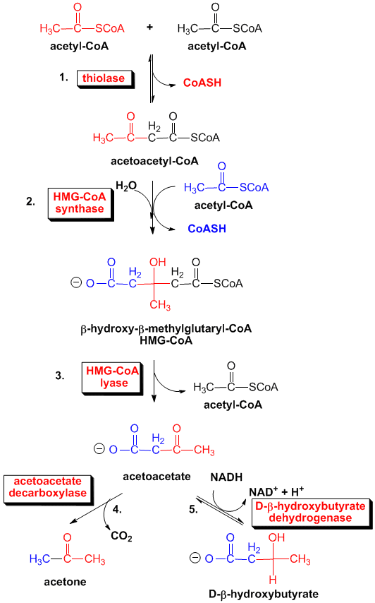
Ketone bodies and it's measurement


TESTING FOR KETONES

The three ketone bodies are acetone (2%), acetoacetic acid (20%) and 3-β-hydroxybutyrate (78%). The primary substrates for ketone body formation are free fatty acids from adipose stores. When glucose is not available ketone bodies supply the majority of the brains energy. After 3 day fast, ketone bodies provide 30% to 40% of the body’s energy requirements. In uncontrolled diabetes the low insulin concentrations result in increased lipolysis and decreased reesterification, thereby increasing plasma free fatty acids. In addition, the increased glucagon: insulin ratio enhances fatty acid oxidation in
Fig. Ketone bodies Synthesis
the liver (as seen in type 1 diabetes). Increased counter-regulatory hormones also augment lipolysis and ketogenesis in adipose tissue and liver respectively. Thus increased hepatic ketone production and decreased peripheral tissue metabolism lead to acetoacetate accumulation in the blood. A major fraction is converted to β-hydroxybutyrate. In healthy people beta hydroxybutyrate and acetoacetate are present at equimolar concentrations, here acetone is minor component. In severe diabetes the ratio between beta hydroxybutyrate to acetoacetate may increase to 6: 1 owing to the presence of large concentration of NADH which favors beta hydroxybutyrate production. Measurement of ketone bodies is recommended for patients with type 1 diabetes during acute illness, stress, pregnancy or elevated blood glucose >300 mg/dL or when the patients has signs of ketoacidosis.

None of the commonly used methods for the detection and determination of ketone bodies in serum or urine reacts with all 3 ketone bodies. Gerhardt’s ferric chloride test reacts with acetoacetate only. Nitroprusside test are least 10 times more sensitive to acetoacetate than to acetone and give no reaction at all with beta hydroxybutyrate. So, most of the tests for ketosis essentially detect or measure acetoacetate only, so presence of ketosis may not be detected. Traditional tests for beta hydroxybutyrate are indirect; they require brief boiling of the urine to remove acetone and acetoacetate by evaporation (acetoacetate first break down spontaneously to acetone) followed by gentle oxidation of beta hydroxybutyrate to acetoacetate and acetone with peroxide, ferric ions or dichromate.  The acetoacetate thus formed may be detected with Gerhardt’s test or one of the procedures using Nitroprusside.

Excessive formation of ketone bodies results in increased blood concentrations (ketonemia) and increased excretion in the urine (ketonuria). This process is observed in conditions associated with decreased availability of carbohydrates (like in starvation or frequent vomiting) or decreased use of carbohydrates (like in diabetes mellitus, glycogen storage disease, von Gierke’s disease and alkalosis). The popular high-fat, low-carbohydrate diets are ketogenic and increase ketones in circulation. Diabetes mellitus and alcohol consumption are the most common cause of ketoacidosis in adults. Ingestion of isopropyl alcohol and salicylate poisoning can also produce ketoacidosis. Urine ketones are widely used for monitoring control in patients with type 1 diabetes, pregnancy with preexisting diabetes and GDM.

Although quantitative determination of individual ketone bodies is possible these methods are not used as routine tests. The semiquantitative acetest and ketostix are frequently used but are insensitive to beta hydroxybutyrate so negative Nitroprusside test result does not rule out ketoacidosis.

The test strip contains a mixture of glycine, sodium Nitroprusside, disodium phosphate, and lactose. Acetoacetate or acetone in the presence of glycine forms a lavender-purple complex with Nitroprusside.

Beta hydroxyl butyrate can be determined by using beta hydroxybutyrate dehydrogenase to convert it to acetoacetate with the production of NADH; this NADH is oxidized to NAD in the presence of NBT and enzyme diaphorase and producing reduced NBT which is purple compound whose absorbance can be measured.

Urine testing for ketone bodies are useful for type 1 diabetes mellitus. This testing is important when glucose levels are persistently higher and associated with symptoms compatible with ketoacidosis like nausea, vomiting or abdominal pain. The commercial test uses Nitroprusside-based (Rothras test) reactions to measure acetoacetate. Certain drugs may cause positive results like captopril and levodopa, while ascorbic acid intake may cause false negative results.

BLOOD GLUCOSE MEASUREMENT


Measured in plasma or whole blood. Plasma values are slightly lower than whole blood. Glucose measurement in urine. In urine the blood glucose ranges in around 180 mg/dl. Serum or plasma is the specimen of choice. For urine 24 hr collections can be done and should be preserved by adding 5 mL of glacial acetic acid in vial. Other preservatives lik 5g sodium benzoate can be used.

Hexokinase and glucose oxidase are the two main types of method used to measure glucose in body fluids.

HEXOKINASE METHOD:

(Source: Tietz clinical chemistry, 4th Edition)

The rate of increase in absorbance by NADH is measured at 340 nm. This is the reference method. Specimen blank is used to correct for interferences. The interfering substances are bilirubin, drugs, lipemia, hemolysis. 

GLUCOSE OXIDASE METHOD:

(Source: Tietz clinical chemistry, 4th Edition) 
  
Glucose oxidase is highly specific for beta-D-glucose. About 36% and 64% of glucose in solution are in alpha and beta form respectively. Complete reaction of glucose requires mutarotation of alpha to beta. Reagents contain mutarotase for this. Otherwise extended incubation time allows spontaneous conversion.
In second step various substances, like uric acid, ascorbic acid, bilirubin, hemoglobin, tetracycline, and glutathione inhibit the reaction by competing with the chromogen for hydrogen peroxide producing lower values. Incroporation of potassium ferrocyanide decreases interference by bilirubin. Incorporation of substituted phenol compounds like dimethyl alanine, danisidine, increases the absorptivity and redox reaction. This type of coupled reaction by using substituted benzene compounds to enhance absorptivity and redox system is known as the trinder reaction
Older procedure use quantative Benedict’s test to determine the glucose in blood and are based on its property of acting as reducing agent in hot alkaline solution. In hot alkaline condition the aldehyde group of glucose readily reduces cupric ions to cuprous ions, which form cuprous oxide. Sodium sulphate is used in the reagent to decrease the solubility of oxygen in reagent otherwise this oxygen will again reoxide the cuprous oxide.
In folin wus method proteins are precipitated with tungstic acid and the clear protein free filtrate is used in the reaction with cupric ions.
In somogyi-Nelson method proteins are precipitated by addition of Ba(OH)2 and ZnSO4. Protein is removed as zinc proteinate sulfhydryl compounds as zinc salts and the remaining zn and Ba ions as zinc hydroxide and barium sulfate. Before the advent of enzyme assay, this was the reference method for glucose.
Another test is O-Toulidine test where O-toulidine (aromatic amine) in hot acidic solution will yield a colored compound (due to formation of Schiff base) with an absorbance maxima at 630 nm.

Glucose in urine can be determined by Benedict’s reagent as qualitative method. Other reagent strips are also available and have glucose oxidase in chromogenic assay. These strips has impregnated with glucose oxidase, peroxidase, and the dye o-toulidine, tetramethylbenzidine. The test end of strip is moistened with freshly voided urine and examined after 10 seconds. A blue color develops if glucose is present at 100 gm/dL or more. Some strips have iodine where hydrogen peroxide produced oxidizes iodide to iodine yielding various intensities of brown color corresponding to glucose concentration in urine.

GLUCOSE METERS

A sample of blood from fingerstick but anticoagulated blood is placed on the test pad. The strip is then inserted into the meter. After a fixed period of time the result appears on digital display screen. The meter use reflectance photometry or electrochemistry to measure the rate of reaction. Reflectance photometry measures the amount of light reflected from a test pad containing reagent. In electrochemical systems, the enzymatic reaction in an electrode incorporated on the test strip produces a flow of electrons. The current which is directly proportional to the amount of glucose is converted to a digital readout. The pad contains dry GOD-POD and chromogen.

Many glucose sensors with electrochemical detectors can be implanted intravenously or subcutaneously. Some instruments can be kept on skin which senses the movement of glucose in extra and intravascular space that produces small current and this is sensed. 


Thursday, November 15, 2012

BIOCHEMICAL MEASUREMENTS IN DIABETES MELLITUS


FOR DIAGNOSIS

PRECLINICAL (screening)

·         Immunological markers – ICA, IAA, GAD, Protein tyrosine phosphatase antibodies (IA-2)
·         Genetic markers – (HLA)
·         Insulin secretion – fasting, pulse, response to glucose challenge
·         Blood glucose

ADA recommends screening of first degree relatives of patients with type 1 diabetes by measuring immune related markers (autoantibodies) to protract the clinical outcome by giving immune intervention.

ADA also advocates screening in all asymptomatic individuals over the age of 45 years for development of type 2 diabetes. Screening should be performed particularly in overweight individuals by FPG. If results are less than 110 mg/dL testing should be repeated at 3 year intervals. Testing may be considered at a younger age or be carried out more frequently in individuals at increased risk of diabetes. For high risk groups testing should be done every 2 years starting at 10 years of age. Rationale for screening is that about 33% of individuals with type 2 diabetes are undiagnosed.

CLINICAL

·         Blood glucose
·         OGTT
·         Ketone (urine and blood)
·         Other (e.g. insulin, C-peptide and stimulation tests)
These tests are done to diagnose diabetes and classification after onset of clinical outcome.

FOR MANAGEMENT

ACUTE

·         Glucose (blood, urine)
·         Ketone (blood, urine)
·         Acid-base status (pH, bicarbonate)
·         Lactate

CHRONIC

·         Glucose (blood, urine)
·         Glycated protein (GHb, Fructosamine)
·         Urinary protein (UAE, microalbuminuria, proteinuria)
·         Evaluation of complications (e.g. creatinine, cholesterol, and Tg)

PATHOGENESIS OF DIABETES MELLITUS: MAIN HYPOTHESIS EXPLAINED


Four main hypotheses have been proposed to explain how hyperglycemia causes the neural and vascular pathology. These are;

a.      Increased aldose reductase (or polyol pathway) flux
b.      Enhanced formation of advanced glycation end products (AGE)
c.       Activation of protein kinase C
d.      Increased hexosamine pathway flux
e.      Generation of ROS and oxidative stress (other pathway)


ACTIVATION OF POLYOL PATHWAY

Here the enzyme aldose reductase involved in reduction of toxic aldehydes to their alcohols is diverted to reduce excess glucose (during intracellular hyperglycemia) to sorbitol which is further oxidized to fructose. This process consumes NADPH, which is not available for regeneration of reduced glutathione, thus rendering cells vulnerable to the effects of oxidative stress. Sorbitol accumulation in the lens may cause cataracts, or in nerves and renal glomeruli leading to retinopathy and nephropathy.

ACCUMULATION OF ADVANCED GLYCATION END PRODUCTS (AGES)


These are irreversibly formed by non-enzymatic glycosylation of matrix, cellular and plasma proteins. Glucose and amino acid combine to form unstable Schiff base adducts, which undergo chemical rearrangement over time to form Amadori products and eventually to stable AGEs which are irreversibly attached to proteins and these can trap other protein elements by covalent binding and promotion of cross linking These products can cause tissue damage by alterations in the structure and function of extracellular matrix, by activation of inflammatory cytokines, by alteration of cellular genetic material. This contributes to endothelial dysfunction, basement membrane thickening and increased vascular permeability.

INTRACELLULAR HYPERGLYCAEMIA DUE TO INCREASED DAG CONCENTRATION


This DAG activates NFkB pathway via activation of protein kinase C (PKC). This leads to vasoconstriction, hypercoagulability via increased endothelin-1, TGF-β and plasminogen activator inhibitor (PAI)-1 generation and reduced eNOS (endothelial nitric oxide synthase) generation.

SHUNTING OF EXCESS INTRACELLULAR GLUCOSE TO HEXOSAMINE PATHWAY


Glucose is shunted to hexosamine pathway via fructose 6-phosphate to glucosamine 6-phosphate, catalyzed by enzyme glutamine: fructose 6-phosphate amidotransferase. Glucosamine 6-phosphage converts to UDP N-acetyl glucosamine which by binding to serine and threonine residues on transcription factors, leads to increased pro-inflammatory cytokine activity (TGF- β, PAI-1 and others)

GENERATION OF ROS AND OXIDATIVE STRESS


Excess intracellular glucose or NEFA stimulate TCA by substrate accumulation. Citrate, formed from NEFA or glucose-derived acetyl-CoA and oxaloacetate, is converted to isocitrate by enzyme isocitrate dehydrogenase generating mitochondrial NADH. Excessive generation of NADH leads to ROS formation via increased ETC along inner mitochondrial membrane. Beta cells are more vulnerable this oxidative stress as they contain less antioxidants. PKC can also be activated by superoxide ions and AGE formation thus linking the oxidative stress, AGE and PKC pathway.
Related Posts Plugin for WordPress, Blogger...