Showing posts with label Diabetes. Show all posts

Tuesday, December 16, 2014

Low blood sugar: What are the effects in our body


Fig. Effects of low blood sugar
Every cell in your body needs sugar (glucose) to function. When your blood sugar levels drop too low, your cells become starved for energy. Initially, that can cause minor symptoms, but if you don’t get your blood sugar levels up soon, you’re at risk of serious complications.

Effects of Low Blood Sugar on the Body

When your blood sugar (glucose) levels fall below the normal range, it’s called hypoglycemia, or insulin shock.

Low blood sugar can happen when you skip a meal. It can also happen if your pancreas releases more insulin than it should after you’ve eaten. The most common reason for low blood sugar is diabetes. In type 1 diabetes, the pancreas can no longer produce insulin. In type 2 diabetes, the pancreas doesn’t make enough, or your body can’t use it properly. To keep blood sugar levels from rising too much (hyperglycemia), you need the right amount of insulin. With
insufficient insulin, your blood sugar levels rise. Too much, and your blood sugar levels can plummet.

Thursday, August 21, 2014

Diabetes could be cured: A study from scientists at Manchester University


Scientists at Manchester University have discovered that the hormone amylin can clump together and stop cells producing insulin

Diabetes could be cured with medication after scientists discovered that clumps of amylin may be stopping cells producing insulin
Fig. Diabetes could be cured with medication after scientists discovered that clumps of amylin may be stopping cells producing insulin (Photo courtesy: Alamy)
Diabetes could be cured after scientists discovered that toxic clumps of a hormone stop cells producing insulin.

Scientists at Manchester University have found that both Type 1 and Type 2 diabetes are driven by the same underlying mechanism.

Monday, June 30, 2014

Gestational diabetes mellitus (GDM): Glucose Challenge Test or Glucose Tolerance Test (GTT)


What is gestational diabetes?
The hormone insulin moves glucose or sugar from your blood and into your body’s cells, where it is used for energy. When you have diabetes, this process is blocked and your cells become “insulin resistant”.  This causes you to have too much glucose in your blood. In pregnancy, the hormones from the placenta, which help your baby to grow, can cause your cells to become insulin resistant. Usually in pregnancy the body produces more insulin to counter this but in some mothers this doesn’t happen and they develop gestational diabetes.

Thursday, June 26, 2014

Raises Tenfold risk by Diabetes gene : A study on Greenland


Fig. Greenland: The island's population is only 57,000
A genetic susceptibility that gives a tenfold increased risk of developing type 2 diabetes has been discovered.
The gene mutation, found in the population of Greenland, will give clues to the different causes of the condition, say Danish scientists.
The research, published in Nature, adds to evidence genetics plays a role in the chances of developing diabetes.

Monday, April 28, 2014

Tanzeum Approved for Type 2 Diabetes

Tanzeum (albiglutide) has been approved by the U.S. Food and Drug Administration to help control blood sugar in adults with type 2 diabetes.


Some 24 million people in the United States have type 2 diabetes, which is more than 90 percent of all diabetes cases, the agency said Tuesday in a news release. People with the disease are at increased risk of heart problems, blindness, nerve damage and kidney damage.


Tanzeum is a glucagon-like peptide-1 receptor agonist, a hormone that helps regulate blood sugar. The injectable drug's safety and effectiveness -- in combination with diet and exercise -- were evaluated in a clinical trial of more than 2,000 people. The drug can be used as a standalone therapy or in combination with other diabetes drugs such as metformin, glimepiride (Amaryl), pioglitazone (Actos) or insulin, the FDA said.


The most common side effects of Tanzeum's use were diarrhea, nausea and injection-site reactions. The drug's label will include a boxed warning that it may increase the risk of thyroid tumors, although a scientific link between the drug and such tumors hasn't been established, the agency said.


The FDA is requiring several post-approval studies to evaluate the drug's possible link to thyroid tumors, its effectiveness among children and any risks to the cardiovascular system. It is not to be used by people with type 1 diabetes, the agency added.


Tanzeum is produced by GlaxoSmithKline, in Wilmington, Del.


View the original article here

Thursday, April 17, 2014

Diabetes complications are falling while number of cases continues to rise

Two reports released this week shed light on the current state of type 2 diabetes in this country, and their conclusions are both promising and sobering. First, the good news: An article in The New England Journal of Medicine shows that rates of diabetes-related problems like heart attack, stroke, and lower-limb amputation are down by more than 50% over the last two decades.
Now the bad news: during the same time period, the number of people with diabetes has soared, according to a report in the Annals of Internal Medicine. In the early 1990s, 5.5% of adults had diabetes. By 2010, the number had nearly doubled, to 9.3%. That translates into about 21 million American adults living with diabetes.
Although Americans are doing a better job of controlling their diabetes, they’re falling far short at preventing it. “The major reason why the prevalence of diabetes is going up is an increase in the number of people who are overweight and obese,” explains Dr. Osama Hamdy, medical director of the Obesity Clinical Program at the Harvard-affiliated Joslin Diabetes Center and author of The Diabetes Breakthrough, a newly published book from Harvard Health Publications.
Nearly 70% of Americans are overweight or obese. And unless something is done to reverse this trend, millions more could edge closer to diabetes.
To describe how being overweight contributes to type 2 diabetes, Dr. Hamdy uses an automotive analogy. “You have more tissue in your body, so you require more glucose [sugar]. Just as if you have a bigger car, you need more gas,” he says. Insulin—the hormone that moves glucose from the blood into the tissues for use and storage—is like the key to that car.
The more sugar that enters the blood, the more insulin is needed to drive it to its intended destination. The pancreas can pump out extra insulin to meet the need, but over time, the body becomes increasingly less sensitive to it and the pancreas eventually reaches its insulin-producing limit. “Then you get diagnosed with type 2 diabetes,” Dr. Hamdy says.
The upside is that the process can be reversed. When you lose weight, you have less tissue, which means you need less sugar—and therefore less insulin. “You become more efficient, like a hybrid car,” says Dr. Hamdy.
In his own research, he’s found that losing just 7% of your body weight can improve insulin sensitivity by about 57%—more than diabetes drugs can usually do. “What we have seen is that once people start to lose weight in the very early stages of diabetes, they can actually reverse the entire course of the disease,” Dr. Hamdy says.
The ability to reverse diabetes might seem dramatic, but the lifestyle changes needed to achieve it aren’t. The cornerstones of diabetes prevention—diet and exercise—can be easily incorporated into even the most hectic schedule.
Take exercise, for example. “Most people don’t know that if you do short bouts of exercise it is even more efficient than if you do longer bouts,” says Dr. Hamdy. People who can’t carve out time to get to the gym could actually see greater results just by incorporating three 10-minute exercise sessions into their everyday routine.
Here’s a sample schedule:
In the morning when you wake up, stretch for 10 minutes.After you eat lunch, take a brisk 10-minute walk.After dinner, strength train with light hand weights or an exercise band for 10 minutes.Those three 10-minute sessions provide stretching, aerobics, and strength training—all essential components of a well-rounded workout. And if you do them every day, they add up to 210 minutes of activity a week.
An unhealthy diet can also be transformed with a couple of basic fixes. One is to divide your plate into three sections:
Fill one with lean protein—fish, legumes, beans, tofu, or skinless chickenFill another with green vegetables—spinach, broccoli, kale, or Brussels sproutsFill the third with whole grains—whole-wheat bread, brown rice, quinoa, etc.The other part of the diet fix is learning how to listen to hunger and satisfaction cues to control portion sizes. Dr. Hamdy recommends assessing your hunger based on a five-point scale:
StarvingHungryOk—satisfiedFullStuffedThe goal should be to sit down to a meal when you’re hungry, and stop eating when you’re satisfied.
Preventing diabetes can be done, but it takes a real commitment to change. And until more people are willing to change their eating and exercise habits—and pass those habits on to their children—the obesity and diabetes rates will continue to rise.


View the original article here

Saturday, April 12, 2014

Foot and ankle exercises in patients with diabetes


By Pamela D. Ritzline, PT, EdD, and Audrey Zucker-Levin, PT, PhD.


Guidelines recommend cardiovascular and strengthening exercises in patients with diabetes, but flexibility exercises focused on the foot and ankle can impart added benefits. Improving range of motion can positively affect gait, pressure distribution, and risk of foot ulceration.


Diabetes mellitus (DM) is a chronic, systemic disorder that disturbs the body’s insulin mechanisms, altering blood glucose levels, which can lead to severe health problems and disability.1 DM is epidemic worldwide with a significant number of people in the United States having this condition. The American Diabetes Association (ADA) reports 23.6 million children and adults in the U.S. have diabetes, with 17.9 million diagnosed, 5.7 million undiagnosed, 57 million in a pre-diabetes state, and 1.6 million new cases diagnosed annually in persons 20 years of age and older (most recent data gathered in 2007). DM is the seventh leading cause of death in the U.S., contributing to 233,619 deaths in 2005 (last year data available). The cost of diabetes care was $174 billion in 2007.2 The number of individuals affected by this disease continues to rise; therefore, holistic care is imperative to control the functional limitations affecting patients with DM.


As the incidence of DM rises, healthcare professionals must recognize the risk factors contributing to the development of the disorder. The list is quite lengthy; however, the common risk factors include obesity, physical inactivity, elevated blood glucose, hypertension (> 140/90), smoking, family history, and abnormal lipid metabolism. The incidence of DM increases with age with men having a slightly greater risk than women, and African Americans having the greatest risk of developing DM.2 The more risk factors a person has, the greater the risk of developing type 2 diabetes and associated medical problems. Associated medical problems include cardiovascular disease, peripheral neuropathy, retinopathy, renal failure, dental disease, erectile dysfunction, ketoacidosis, hyperlipidemia, cognitive impairment, an increased susceptibility to other illnesses such as pneumonia or influenza, decreased range of motion of the feet and ankles, balance impairment, and non healing ulcers that may lead to amputation.2


Exercise and diabetes


The benefits of exercise for the diabetic population are widely described in the literature. Although a detailed discussion of the evidence supporting the need for exercise in persons with type 2 diabetes is beyond the scope of this article, a preponderance of such evidence exists.2-18 A Pubmed/Medline search using the words “diabetes, exercise, training” yielded 2481 citations. Culling through the citations revealed studies with outcomes that supported exercise for improving VO2max anaerobic threshold, time to anaerobic threshold,3 improving endurance,3,4 improving strength,5-8 improving metabolic control,9-12 improving emotional well being,8,9 and improving mental health and vitality8 while decreasing metabolic syndrome risk factors13 decreasing insulin requirement,9,11 and decreasing falls.14,15 Exercise also has been shown to increase the cells’ sensitivity to insulin, improve blood glucose control (decreasing the amount of medications necessary)2 decrease hypertension, improve lipid metabolism leading to a healthier heart, assist with weight control, reduce cardiometabolic risks, improves sleep patterns and energy levels, reduce stress, increase flexibility, and build stronger bones and muscles.2,16-18 No negative effects from exercise were discovered in the citations reviewed.


The extensive literature review revealed that exercise interventions varied in longevity (from one week to a lifetime), in duration (from two to seven days per week), and in intensity. Some interventions focused on cardiovascular fitness, others on muscle strengthening, balance, and/or flexibility. Some programs incorporated multiple interventions. So, which exercises are most beneficial and how do we assure compliance long term?


Five basic categories of exercise are recognized: cardiovascular, strength, flexibility, balance, and cognitive. The U.S. federal government has published Physical Activity Guidelines for all Americans, including those with chronic disease such as diabetes.1 The guidelines clearly state that adults should participate in a total of 150 minutes of moderate-intensity or 75 minutes of vigorous-intensity aerobic physical activity per week. Exercise should be spread over the entire week with sessions divided into 10 minute intervals. The simplest exercise would be to encourage individuals to walk at a vigorous pace while wearing appropriate footwear. Other options involve use of equipment such as a bicycle, elliptical, stepper, or treadmill to accomplish the cardiovascular requirement.1


The guidelines also recommend that adults perform at least twice weekly strengthening exercises of all major muscle groups. People can be instructed in exercises that use bodyweight as resistance or machines for resistance. Major muscle groups include the quadriceps, hamstrings, calves, abdominals, biceps, triceps, and forearms. Guidelines vary on intensity and frequency, with benefits reported from performing one set of 15 moderate intensity repetitions to three sets of 15 repetitions of low intensity repetitions. The frequency and intensity should be tailored based on an individual’s perceived ability to maintain the program.1


In addition to cardiovascular and strengthening exercise, people with DM should participate in a flexibility program. Peripheral neuropathy, as a result of diabetes, may cause sensory and proprioceptive loss in the extremities and decreased range of motion, specifically at the ankles, feet, and shoulders. Zimny et al reported progressive stiffening of collagen containing tissue in people with DM, which may add to joint stiffness.19 This increased joint stiffness results in diminished range of motion, but because the associated clinical disability is often subtle, it is often overlooked.19


Decreased range of motion in the ankle and foot may cause gait abnormalities, such as a footflat contact or a forefoot contact gait pattern. Either pattern eliminates the energy conserving and force attenuating motion that occurs at the ankle and subtalar joint during a normal gait cycle. Further, diminished metatarsophalangeal joint dorsiflexion in the pre-swing phase of gait may result in decreased balance and abnormal forefoot pressure. If MTP joint dorsiflexion is not sufficient, the person may shorten their step length and exert more energy to lift the leg from the support surface because a rigid lever for push-off is not achieved. This gait pattern may increase susceptibility to falls and injury. In addition to balance impairment, joint stiffness throughout the foot may lead to increased midfoot and forefoot pressure when walking. This can increase the risk of ulceration, particularly in patients with peripheral neuropathy.20 This is supported by the findings of Fernando et al, who reported that limitations in subtalar and first metatarsophalangeal joint mobility resulted in increased peak foot pressures during gait.21


Foot and ankle exercises


Goldsmith et al found that persons with DM who were educated in a foot and ankle exercise program experienced decreased joint stiffness and decreased peak plantar pressure during gait after only one month of intervention.22 The regimen included “drawing” the alphabet with the foot (ABCs), passive and active dorsiflexion and plantarflexion of the metatarsophalangeal joint, passive and active dorsiflexion and plantarflexion of the ankles, active pronation and supination of the subtalar joint, stretching of the gastrocnemius and soleus, followed by soft tissue manipulation of the entire foot. Exercise may improve joint stiffness and impact ulcer healing. Flahr evaluated the effect on neuropathic foot wounds of non-weightbearing foot and ankle exercises (including active inversion, eversion, dorsiflexion and plantarflexion) performed 10 times each twice a day, and found that those patients who participated in the exercise program trended toward more rapid healing. Flahr attributed the healing to improved blood supply in the area.23


We performed a randomized, controlled study on the effect of a six-week home exercise program, which focused on range of motion of the first metatarsophalangeal joint and the talocural joint, on gait parameters in persons with type 2 DM.24 Ten subjects participated in a home exercise program (HEP) developed according to the American College of Sports Medicine guidelines. The HEP consisted of a five to 10 minute warm-up walk, ABCs, heel raises, toe raises, and towel exercises (dorsiflexion, eversion, and inversion) to be performed three times per week for six weeks. A difference in plantarflexion ROM was the only significant effect of the exercise program for our population. No significant changes in gait were noted. This may have resulted from the short duration of the program.


Patients diagnosed with DM should be instructed in a home exercise program that focuses on maintaining or improving range of motion in the ankle and foot. Simply “drawing” an exaggerated alphabet with the ankle twice daily; actively performing ankle dorsiflexion, plantarflexion, inversion, eversion 10 times each twice daily; and manually mobilizing the forefoot, including the first metatarsophalangeal joint, into dorsiflexion will help to increase range of motion of the foot, diminish peak foot pressure, and possibly prevent breakdown.


Balance exercises also should be incorporated into the daily exercise regime of someone with diabetes. Wrobel and Najafi25 reported that people with diabetes walk with “a conservative gait strategy” characterized by a wider base of support and prolonged double support time. This gait pattern may be a protective strategy to counter the effects of decreased balance from diminished proprioception. For this reason, daily balance exercises in a protected environment are recommended. Persons with diabetes can be encouraged to maintain their balance by standing on one foot while brushing their teeth or while washing the dishes. Balance activities can be progressed to be more challenging, first by performing dynamic activities such as raising the arms or catching a ball while standing on a wide stable base of support with two feet on the ground, then by performing the same activity with a progressively smaller base of support, such as standing on one foot. Balance activities can be progressed from a stable surface to unstable surfaces such as sand or a dome while diminishing the base of support area.


Cognitive exercise is a relatively new area of study. Van Elderen et al found increased progression of brain atrophy and decreased cognitive function in patients with diabetes compared to normal controls.26 Although data do not exist to support the benefits of cognitive exercise in people with diabetes, initiating a cognitive exercise program may help maintain cognitive function. Spending 30 minutes daily learning a new skill, such as a foreign language or an instrument, or even doing daily crossword puzzles or other brain teasers may help maintain or improve cognitive function in people with diabetes.


Adherence to an exercise program is challenging for any individual, independent of diabetes. Beverly and Wray27 reported that collective support, motivation and responsibility all play a role in empowering an individual to stick with an exercise program.


Lastly, before engaging in any exercise program, patients with diabetes should consult a physician to ensure participation in such activity is safe. Once the patient has been cleared to exercise, a physical therapist can design an individualized program that incorporates the appropriate exercises and focuses on the needs of the patient. Individuals with DM must be cautioned to monitor blood glucose levels before, during, and after exercise to avoid a hypoglycemic event during or after exercise as well as a hyperglycemic event 24 hours post exercise.2,16,17 As health care providers, we must support and encourage exercise in our patients by being role models and advocates.


DM is epidemic in the U.S. Maintaining health through exercise decreases costs and improves quality of life.28 A preponderance of evidence suggests that every person with DM should participate in a consistent exercise program. We, as health care providers, must work together to identify patients in need of guidance and direct them to the appropriate practitioners who understand the potential complications unique to this population.


Pamela D. Ritzline, PT, EdD, is associate professor and director of the graduate program in the department of physical therapy at the University of Tennessee Health Science Center in Memphis. Audrey Zucker-Levin, PT, PhD is an associate professor in the same department.


References:


1. CDC National Diabetes Fact Sheet US, 2005. Available at: http://www.cdc.gov/diabetes/pubs/pdf/ndfs_2005.pdf


2. American Diabetes Association. Diabetes statistics. Available at: http://www.diabetes.org/diabetes-basics/diabetes-statistics/


3. Mourot L, Boussuges A, Maunier S, et al. Cardiovascular rehabilitation in patients with diabetes. J Cardiopulm Rehabil Prev 2010;30(3):157-164.


4. Pariser G, DeMeuro M,Gillette P, Stephen W. Outcomes of an education and exercise program for adults with type 2 diabetes, and comorbidities that limit their mobility: a preliminary project report. Cardiopulm Phys Ther J 2010;21(2):5-12.


5. Kwon HR, Han KA, Ku YH, et al. The effects of resistance training on muscle and body fat mass and muscle strength in type 2 diabetic women. Korean Diabetes J 2010;34(2):101-110.


6. Plotnikoff RC, Eves N, Jung M, et al. Multicomponent, home-based resistance training for obese adults with type 2 diabetes: a randomized controlled trial. Int J Obes 2010;34(12):1733-1741.


7. Larose J, Sigal RJ, Boule NG, et al. The effect of exercise training on physical fitness in type 2 diabetes mellitus. Med Sci Sports Exerc 2010 Feb 4. [Epub ahead of print]


8. Aylin K, Arzu D, Sabri S, et al. The effect of combined resistance and home-based walking exercise in type 2 diabetes patients. Int J Diabetes Dev Ctries 2009;29(4):159-165.


9. Shenoy S, Guglani R, Sandhu JS. Effectiveness of an aerobic walking program using heart rate monitor and pedometer on the parameters of diabetes control in Asian Indians with type 2 diabetes. Prim Care Diabetes 2010;4(1):41-45.


10. Kirwan JP, Solomon TP, Wojta DM, et al. Effects of 7 days of exercise training on insulin sensitivity and responsiveness in type 2 diabetes mellitus. Am J Physiol Endocrinol Metab 2009;297(1):E151-E156.


11. Biesenbach G, Bodlaj G, Sedlak M, et al. Exercise program for older patients with insulin-treated type 2 diabetes: long-term effects on metabolic control and BMI. Z Gerontol Geriatr 2009;42(6):465-469.


12. Zanuso S, Jimenez A, Pugliese G, et al. Exercise for the management of type 2 diabetes: a review of the evidence. Acta Diabetol 2010;47(1):15-22.


13. Strasser B, Siebert U, Schobersberger W. Resistance training in the treatment of the metabolic syndrome: a systematic review and meta-analysis of the effect of resistance training on metabolic clustering in patients with abnormal glucose metabolism. Sports Med 2010;40(5):397-415.


14. Kruse RL, Lemaster JW, Madsen RW. Fall and balance outcomes after an intervention to promote leg strength, balance, and walking in people with diabetic peripheral neuropathy: “Feet First” randomized controlled trial. Phys Ther 2010;90(11):1568-1579.


15. Morrison S, Colberg SR, Mariano M, et al. Balance training reduces falls risk in older individuals with type 2 diabetes. Diabetes Care 2010;33(4):748-750.


16. Manders RJ, Van Dijk JW, van Loon LJ. Low-intensity exercise reduces the prevalence of hyperglycemia in type 2 diabetes. Med Sci Sports Exerc 2010;42(2):219-225.


17. Goodman CC, Boissonnault WG, Fuller KS. Pathology: Implications for the Physical Therapist. 3rd ed. St. Louis: Saunders Elsevier; 2008.


18. Marwick TH, Hordern MD, Miller T, et al. Exercise training for type 2 diabetes mellitus: impact on cardiovascular risk: a scientific statement from the American Heart Association. Circulation 2009;119(25):3244-3262.


19. Zimny S, Schatz H, Pfohl M. The role of limited joint mobility in diabetic patients with an at-risk foot. Diabetes Care 2004;27(4):942-946.


20. Sacco IC, Hamamoto AN, Gomes AA, et al. Role of ankle mobility in foot rollover during gait in individuals with diabetic neuropathy. Clin Biomech 2009;24(8):687-692.


21. Fernando DJ, Masson EA, Veves A, Boulton AJ. Relationship of limited joint mobility to abnormal foot pressures and diabetic foot ulceration. Diabetes Care 1991;14(1):8-11.


22. Goldsmith JR, Lidtke RH, Shott S. The effects of range-of-motion therapy on the plantar pressures of patients with diabetes mellitus. J Am Podiatr Med Assoc 2002;92(9):483-490.


23. Flahr D. The effect of nonweight-bearing exercise and protocol adherence on diabetic foot ulcer healing: a pilot study. Ostomy Wound Manage 2010;56(10):40-50.


24. Ritzline PD, Swanson J. The effects of a home exercise program on ankle range of motion and step length in people with type 2 diabetes: A blinded cross-over design pilot study. Presented at the annual meeting of the American Physical Therapy Association, Boston, June 2010.


25. Wrobel JS, Najafi B. Diabetic foot biomechanics and gait dysfunction. J Diabetes Sci Technol 2010;4(4):833-845.


26. van Elderen SG, de Roos A, de Craen AJ, et al. Progression of brain atrophy and cognitive decline in diabetes mellitus: a 3-year follow-up. Neurology 2010;75(11):997-1002.


27. Beverly EA, Wray LA. The role of collective efficacy in exercise adherence: a qualitative study of spousal support and type 2 diabetes management. Health Educ Res 2010;25(2):211-223.


28. Sacks N, Cabral H, Kazis LE, et al. A web-based nutrition program reduces health care costs in employees with cardiac risk factors: before and after cost analysis. J Med Internet Res 2009;11(4):e43.


Friday, April 11, 2014

Elderly Diabetes Patients on Insulin Most Vulnerable to Low-Blood-Sugar Trouble

By Serena Gordon

A new look at diabetes patients in the United States who use insulin and wind up in the emergency room with low blood sugar shows the dangerous scenario is more than twice as likely to happen to those over 80 years old.

Diabetes in Middle Age May Cause Memory Problems Later

By Dennis Thompson
HealthDay Reporter

People who develop type 2 diabetes or high blood pressure in middle age appear more likely to suffer brain damage that can contribute to dementia as they grow older, a new study finds.


Diabetes might actually shrink the brain over a long period of time, reducing the size of crucial areas like the hippocampus, which plays an important role in short- and long-term memory, according to the study.


Additionally, diabetes and high blood pressure both seem to increase a person's risk of micro-strokes and other damage to the blood vessels that feed the brain, the study authors said.


"People who had diabetes earlier in life had much worse brain [structure] than those who had it later in life," said lead author Dr. Rosebud Roberts, a Mayo Clinic researcher. "These scans are showing us that cognitive impairment happens over a long period of time. The earlier you develop type 2 diabetes, the more likely you are to have damage."


Diabetes has long been linked to problems with thinking and memory later in life, but this study is the first to provide solid evidence explaining why that occurs, said Keith Fargo, director of scientific programs and outreach for the Alzheimer's Association.


"We are very excited about this study," Fargo said. "It has been known for quite some time that there is some kind of link between diabetes and cognitive ability later in life. What has not been known yet is why this link exists and how it develops over time."


The study involved more than 1,400 people with an average age of 80, according to the report published online March 19 in the journal Neurology. The study participants had at most slight memory and thinking problems called mild cognitive impairment. The researchers assessed the participants' thinking and memory skills, noting any signs of mild impairment.


The study participants then underwent MRI brain scans to look for signs of brain damage that can be an early indication of dementia.


Finally, the researchers reviewed the participants' medical records to see whether they had been diagnosed with diabetes or high blood pressure in middle age, which, for the purposes of this study, ran from 40 to 64.


The study authors found that people who developed diabetes in middle age had brains that were on average 2.9 percent smaller than people who didn't have diabetes. And their hippocampi were even smaller -- an average of 4 percent smaller than those of non-diabetics.


"When your hippocampus begins to shrink, you begin to lose your long-term memory and your ability to remember recent events," said Roberts, who also is a member of the American Academy of Neurology.


Midlife diabetes also was associated with an 85 percent greater risk of micro-strokes in the brain. Finally, people with middle-age diabetes were twice as likely to have thinking or memory problems, the study found.


And people with high blood pressure in midlife were twice as likely to have damage caused by stroke to portions of the brain associated with thought, memory and language, the researchers said.


Although the study uncovered an apparent link between diabetes or high blood pressure in middle age and memory problems later in life, it didn't prove a cause-and-effect relationship.


The research results emphasize the need for people to adopt a healthy lifestyle in middle age or earlier, Fargo said.


Recent polls show that nearly a quarter of people mistakenly think they're at risk for Alzheimer's disease only if it runs in their family, he said. In reality, dementia can strike anyone if they don't take good care of themselves, he added.


"If you've got a brain, you're at risk for dementia," Fargo said. "Midlife is really going to be a critical time for people to focus on their brain health, and not wait until it's too late."


People who want to protect their brain health should avoid developing diabetes or high blood pressure, Roberts said. She noted that even people who became diabetic in old age still suffered areas of brain damage as a result of the disease.


If a person does develop either chronic condition, they can limit the impact on thinking and memory by controlling the disease with diet, exercise and medication, the researchers said.


"If you have type 2 diabetes, you have an increased risk of brain damage," Roberts said. "But if you control your diabetes well, it should reduce the damage that is being caused in your brain."

Thursday, April 10, 2014

Take Heart: Mediterranean Diet Combats Diabetes, Study Says

Adhering to a so-called Mediterranean diet may reduce your risk of diabetes, especially if you're at high risk for heart disease.

That's the finding of researchers who reviewed 19 studies that included more than 162,000 people in different countries for an average of 5.5 years.

Diabetes Tied to Higher Risk of Pancreatic Cancer in Study


By Serena Gordon

People with DM have double the risk of pancreatic cancer compared to people who don't have diabetes, according to a new analysis of 88 previous studies.

Sunday, June 9, 2013

Relation between HbA1c and Estimated Average Glucose (eAG)


Why is relating HbA1c to glucose important?

We are frequently asked about the relationship between HbA1c and plasma glucose levels. Many patients with diabetes mellitus now perform self-monitoring of blood glucose (SMBG) in the home setting, and understanding the relationship between HbA1c and glucose can be useful in setting goals for day-to-day testing.

Thursday, June 6, 2013

Hormones and Body Weight

Excessive intake of calories in relation to energy expenditure over a long period of time results in body weight gain.

A complex physiologic system regulates energy homeostasis by integrating signals from peripheral organs with central coordination in the brain. The hypothalamus functions as the main cerebral center in which these signals converge.

Monday, November 19, 2012

GLYCOGEN STORAGE DISEASE


Glycogen storage diseases are the result of deficiency of enzymes that cause the alteration of glycogen metabolism. The liver forms (type I, III, IV and VI) are marked by hepatomegaly due to increased liver glycogen and hypoglycemia caused by inability to convert glycogen to glucose. The muscle forms (type II, IIIA, V and VII) have mild symptoms appearing during sternous exercise owing to inability to provide energy for muscle contraction.

INBORN ERROR OF CARBOHYDRATE METABOLISM


Deficiency or absence of an enzyme that participate in carbohydrate metabolism may result in accumulation of monosaccharides, which can be measured in urine. Most of these conditions are inherited as autosomal recessive traits.

DISORDER OF GALACATOSE METABOLISM
Galactose is derived from milk in diet. It is the C4 epimer of glucose. A deficiency of any of the enzyme that participates in conversion of galactose to glucose results in galactosemia. Galactosemia occurs due to inhibition of glycogenolysis.

GALACTOSE-1-PHOSPHATE URIDYL TRANSFERASE DEFICIENCY

Infants with this deficiency fail to thrive on milk because half of the milk sugar, lactose is galactose. Within few days of milk ingestion neonates manifest vomiting and diarrhea. Failure to thrive, liver disease, cataracts and mental retardation develop later. This disorder is identified by measuring erythrocyte galactose -1-phosphate uridyltransferase activity.

GALACTOKINASE DEFICIENCY

This is milder condition manifested by cataracts caused by galactitol deposits in the lens. The diagnosis is confirmed by demonstrating normal transferase activity no galactokinase in red blood cells.

DISORDER OF FRUCTOSE METABOLISM
Fructose may appear in the urine after eating fruits, honey, and syrups, but has no significance in these conditions. Three disorders of fructose metabolism inherited as autosomal recessive trait produces fructosuria.

Essential fructosuria
This occurs due to deficiency of fructokinase

Hereditary fructose intolerance

A deficiency of fructose-1-phosphate aldolase produces this disorder with hypoglycemia and liver failure. Fructose ingestion inhibits glycogenolysis and gluconeogenesis, producing hypoglycemia.

Hereditary fructose-1, 6-diphosphate deficiency

DISORDER OF PENTOSE METABOLISM

Alimentary pentosuria
Pentose may be present in the urine after eating large quantities of fruits such as cherries, plums, or prunes.

Essential pentosuria
This is harmless inborn error caused by deficiency of L-xylulose reductase an enzyme involved in the glucuronic acid pathway.
Individual sugars can be measured by qualitative tests and chromatography

Emergency treatment of hypoglycemia


Glucose should be administered orally (10-20 g in adult patient, 3 times before giving a meal). If oral therapy is not possible the parenteral dose of glucose for adult should be 25-50g as 50-100 mL of 50% dextrose should be given. Failure to respond to glucose, glucagon should be administered intramuscularly or intravenously, steroids (hydrocortisone).

Causes of hypoglycemia:

1.    Medical therapy of diabetes especially insulin administration or oral hypoglycemia drug is the most common cause of fasting hypoglycaemia.
2.   Surreptitious (self-induced) administration of hypoglycemic agents (factitious or felonious hypoglycaemia) like insulin, sulphonylureas, metiglinides, etc.
3.      Insulinoma: Insulin producing islet cell tumors.
4.      Autoimmune hypoglycemia 

In one condition antibodies binds to insulin receptors and mimic the action of insulin. Laboratory finding shows high plasma insulin concentrations but suppressed C-peptide and proinsulin. The other syndrome, autoimmune insulin syndrome, in which antibodies are direction towards insulin. Laboratory finding shows high plasma concentration of insulin and C-peptide (C-peptide level is quiet less than insulin).

1.      Hypoglycaemia associated with renal failure:

Renal impairment leading to hypoglycaemia is the second most common cause of hypoglycaemia, after insulin therapy. The most important factor here is calorie restriction. In normal subjects, the kidney, by gluconeogenesis supply 45% of glucose during prolonged starvation. In uraemic patient this process is impaired. Other mechanisms include increase insulin half-life due to impaired renal clearance and degradation.
2.      Hypoglycemia associated with liver disease:
Liver can maintain glucose homeostasis even functioning liver mass reduces to <20% and hypoglycemia does not occur unless liver is extensively damaged. Conditions like fatty liver, cirrhosis, infective hepatitis, hepatocellular carcinoma are associated with hypoglycaemia.
3.      Alcohol induced hypoglycaemia: 

Alcohol induced fasting hypoglycemia is due to direction inhibition of gluconeogenesis. This is due to accumulation of NADH and increased NADH/NAD+ ratio resulting from the oxidation of ethanol. Alcohol induced fasting hypoglycemia usually develops 6-36h after ingestion of alcohol. There is severe metabolic acidosis with high blood lactate. Hyperketonaemia and ketonuria are present predominantly β-OHB, since the accumulation of NADH suppress the conversion of it to acetoacetate. Prompt IV glucose treatment should be done.

Alcohol potentiates the hypoglycemic effect of insulin and sulphonylurea drugs. Alcohol potentiates the insulin-stimulating effect of glucose and thus increase the risk of reactive hypoglycemia. This is seen during consumption of alcohol and sucrose (e.g. in syrup or tonic) in empty stomach and followed by not eating for few hours afterward. This effect is not seen when saccharin or fructose is substituted for sucrose as sweetening agent. Starchy foods like breads increase the risk fro reactive hypoglycaemia, whereas foods providing fat or protein have the reverse effect.
During exercise, during the first 5-10 minutes of severe exercise, muscle glycogen is the source of energy, by 40 min, 75-90% of glucose is supplied by blood, mainly from increased hepatic glucose production (75% from glycogenolysis and 25% from gluconeogenesis).
4.      Reactive (alimentary) or postprandial hypoglycemia

This occur after gastric surgery, antibodies to insulin, inborn error of metabolism. Symptoms occurring 2-4h after food ingestion and last for about 10-20 min. This is also seen in patients with hereditary fructose intolerance after ingestion of fructose.

Hypoglycemia in Diabetes Mellitus

Hypoglycemia occur frequently in both type 1 and 2 diabetes. This occurs in diabetic patients using hypoglycemia drugs or insulin. In many patients with type 1 disease do not experience the neurogenic warning symptoms for years and are prone to severe hypoglycemia this is called hypoglycemia unawareness. 

HOW TO IDENTIFY THE CAUSE OF HYPOGLYCEMIA


IDENTIFICATION OF CAUSE OF HYPOGLYCEMIA

PLASMA INSULIN AND C-PEPTIDE

Increase in Insulin and C-peptide in the presence of hypoglycemia indicates islet-cell tumors, autoimmune insulin secretion, and drug-induced (sulphonylureas, repaglinide) causing endogenous hyperinsulinaemia.
Decrease in insulin and C-peptide indicates presence of other secondary conditions like chronic renal failure (as C-peptide is excreted by kidney), liver disease, alcohol induced, anorexia nervosa, etc.
Increase in insulin but decrease in C-peptide indicates administration of exogenous insulin, Insulin anti-receptor antibodies (IR-A).

PLASMA Β-HYDROXYBUTYRATE

Hypoglycemia due to hyerinsulinemia shows low ketone bodies. In hypoglycemia due to other conditions like liver disease, anorexia nervosa, hypopituitarism etc, this ketone body is raised.

PLASMA PROINSULIN

Normally only <20% of insulin is released in circulation. In islet cell tumor, circulating proinsulin is increased.

INSULIN ANTIBODIES

The presence of insulin antibodies, due to pre-exposure to exogenous insulin may give false high plasma insulin concentrations. Since C-peptide does not cross-react with insulin antibodies, its measurement can be used as index of β-cell function. 

How Hypoglycemia is investigated ?

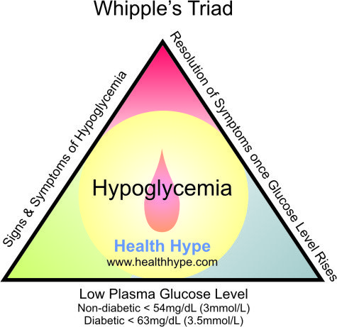
A venous plasma glucose concentration below 50 mg/dl is called hypoglycaemia. The diagnosis of hypoglycemia necessitates the presence of Whipple’s triad. This consists of:

Fig. Whipple's Triad
1)   Symptoms of hypoglycemia
2)   Low plasma glucose concentration and
3)   Symptoms relieved by glucose administration.

Fig. Classical Signs and symptoms of Hypoglycemia
The classic signs and symptoms of hypoglycemia are trembling, sweating, nausea, rapid pulse, lightheadedness, hunger and epigastric discomfort. Neuroglycopenia can be seen in severe cases (headache, confusion, blurred vision, dizziness, and seizures).

The most common cause of hypoglycemia are drugs like propranolol, salicylate, oral hypoglycemic drugs with long half life like chlorpropamide, insulin secreting sulfonylureas, glycogen storage disease, alcoholism, septicemia, hepatic failure, Addison’s disease etc. 

REGULATORY RESPONSE TO HYPOGLYCEMIA

In hypoglycemia, the shortage of glucose in neurons activates hypothalamus, and an autonomic response to restore and maintain glucose supply initiates which has many effects like:

ACTIVATION OF SYMPATHETIC NERVOUS SYSTEM

 α-ADRENERGIC EFFECTS


  • Inhibition of endogenous insulin release
  • Increased cerebral blood flow (peripheral vasoconstriction)

 β-ADRENERGIC EFFECTS:


  • Stimulation of glycogenolysis
  • Stimulation of glucagon release (also α cells can sense directly)
  • Stimulation of lipolysis
  • Inhibition of muscle glucose uptake
  • Increased cerebral blood flow (by increasing cardiac output)

CATECHOLAMINE RELEASE FROM ADRENAL MEDULLA


  • Potentiates the α and β adrenergic effects

ACTIVATION OF PARASYMPATHETIC NERVOUS SYSTEM

  • Stimulates vagus nerve
  • Stimulation of gastric acid secretion
  • Stimulation of parotid salivary secretion. 

There is hierarchy of response of counter-regulatory hormones; glucagon, epinephrine, cortisol and GH. Glucagon and epinephrine are rapidly acting hormones whereas latter two are slow acting and are active at late phase of hypoglycemia. During fast state the first mechanism is inhibition of endogenous insulin secretion and followed by release of counter regulatory hormones in hierarchy.

Decreased endogenous insulin occurs at glucose level 80 mg/dl; increase glucagon, adrenaline, cortisol and GH secretion at 60 mg/dl and development of hypoglycaemic symptoms occurs at 50 mg/dl and impairment of cognitive function at 40 mg/dl. 

INVESTIGATION OF HYPOGLYCEMIA

First is demonstration of hypoglycemia and second to identify the cause of hypoglycemia.

DEMONSTRAITON OF HYPOGLYCEMIA

MEASUREMENT OF BLOOD GLUCOSE
Measurement of blood glucose (insulin, C-peptide) during acute neuroglycopenia (characterized by sweating, anxiety, hunger, palpitation and weakness) is the best test for the diagnosis of hypoglycemia.
PROVOCATION TEST:
Prolonged fast:
This is the single most useful test to evaluate suspected hypoglycemia. The aim of this test is to demonstrate spontaneous hypoglycemia in the presence of neuroglycopenic symptoms during prolonged fasting for 48 h, and that the symptoms resolve on glucose administration.

During the fasting period blood glucose, insulin, C-peptide is measured at every 4-6 hours. But as glucose level falls below 50 mg/dl frequent sample must be taken. About 95% of patient will develop hypoglycaemia within 48 h. Measurement of β-hydroxybutyrate and its raising presence indicates suppression of insulin release and fast can be terminated by giving glucose when FBS becomes <45 mg/dL and patient exhibit signs or symptoms of hypoglycemia.
Mixed meal test:
This is used to investigate patients who experience postprandial symptoms, for the possibility of reactive hypoglycemia. Meal is ingested and plasma glucose measured every 30 min for 6h and at any time during symptomatic phase. Patients developing neuroglycopenia symptoms during hypoglycemia, but not at other times during the test, are considered to have postprandial hypoglycemia. 

COMPLICATIONS OF DIABETES


DIABETIC RENAL DISEASE (DIABETIC NEPHROPATHY)

It is most common in type 1 diabetes. Some 20-30% of patients with type 1 diabetes will develop renal disease (15-25 years after diagnosis). It is less prevalent in type 2 diabetes (only 10-20% lifetime risk).

HYPERFILTRATION AND MICROALBUMINURIA

The earlier symptoms of diabetes includes hyperfiltration (with urine albumin excretion, UAE, <30mg/24 hour or 20µg/min) followed by progression through microalbuminuria to proteinuria (UAE>300 mg/24 h or 200µg/min). After this GFR falls and progress to ESRF. The first and best opportunity to detect the disease clinically is at the stage of microalbuminuria. Dip-stick testing or urine is not usually positive at such concentration of albumin and detection relies on either 24 h quantitation or more conveniently the use of albumin/creatinine ratio (normal <2.5 mg/mmol in men and <3.5 mg/mmol in women) on at least two out of 3 separate urine specimens over a 3-6 month period can be done. Due to day to day variation of UAE rates 2 of 3 samples should be positive for the diagnosis. Microalbuminuria is not just a risk factor of nephropathy but an independent risk factor for CAD (one of the most potent risk factors known), being also associated with dyslipidaemia, hypertension, endothelial dysfunction and diabetic retinopathy.

TYPE 4 RENAL TUBULAR ACIDOSIS

Hyporeninaemic hypoaldosteronism may be a manifestation of diabetic nephropathy. It presents with hyperchloraemic, hyperkalaemic metabolic acidosis. Failure of renin to rise in response to posture or sodium restriction suggest an interstitial (juxtaglomerular) defect. The failure of aldosterone release to be stimulated directly by resulting hyperkalaemia suggest the possibility of dysfunction of adrenal zona glomerulosa.

CHARCOT FOOT

It is a specific foot deformity occurring due to neuropathy and if untreated leads to bone collapse of the foot causing outward bowing. 

OTHER DIABETIC EMERGENCIES: HYPEROSMOLAR HYPERGLYCAEMIC STATES AND ALCOHOLIC KETOACIDOSIS


HYPEROSMOLAR HYPERGLYCAEMIC STATES

Initially called hyperosmolar non-ketotic (HONK) hyperglycaemia. The dominant clinical feature is dehydration. It mainly occurs in older subjects with type 2 diabetes mellitus. The cycle of hyperglycaemia, dehydration (occurring due to vomiting, polyuria, glycosuria osmotically takes more water in urine) and increased counter regulatory hormones (induced by acidosis and dehydration and hyperglycemia) is same in ketoacidosis but is more severe. There is hypernatremia caused by renal sodium resorption in response to hypovolaemia, together with osmotic diuresis causing persistent free water loss. 

Non-ketotic hyperosmolar state usually occurs during marginal insulin deficiency, and their insulinaemia has sufficient antilipolytic effect to prevent the lipolytic and ketotic problems seen in ketoacidosis. There is decrease in anion gap <20 mmol/L and bicarbonate is normal and pH >7.30. There is hypernatraemia and more severe water loss 18 L in typical adult.

ALCOHOLIC KETOACIDOSIS

During alcoholism and resulting poor diet is association with vomiting, this cause ketoacidosis and low, normal or elevated blood glucose. Ketosis is caused by lack of insulin action which results in mobilization of NEFAs and their conversion to ketone bodies as alternative fuel. This is potentiated by counter-regulatory hormones like glucagon, cortisol and catecholamines secreted in response both to hypoglycaemia and extracellular fluid volume contraction. 

In addition, alcohol metabolism depletes cellular NAD+ which by restricting pyruvate formation from lactate, causes accumulation of lactate and depletion of pyruvate, a gluconeogenic substrate. As is the case in DKA, alteration in mitochondrial redox state favors beta hydroxybutyrate over acetoacetate production. A complex acid-base disorder ensues from the combined effects of ketosis causing metabolic acidosis, and a combination of extracellular fluid contraction and vomiting causing metabolic alkalosis. 
Related Posts Plugin for WordPress, Blogger...